A Jaw in Three Planes
A flat X-ray shows a shadow. A cone beam scan shows the teeth, the bone and the joints as a volume you can turn, slice and measure.

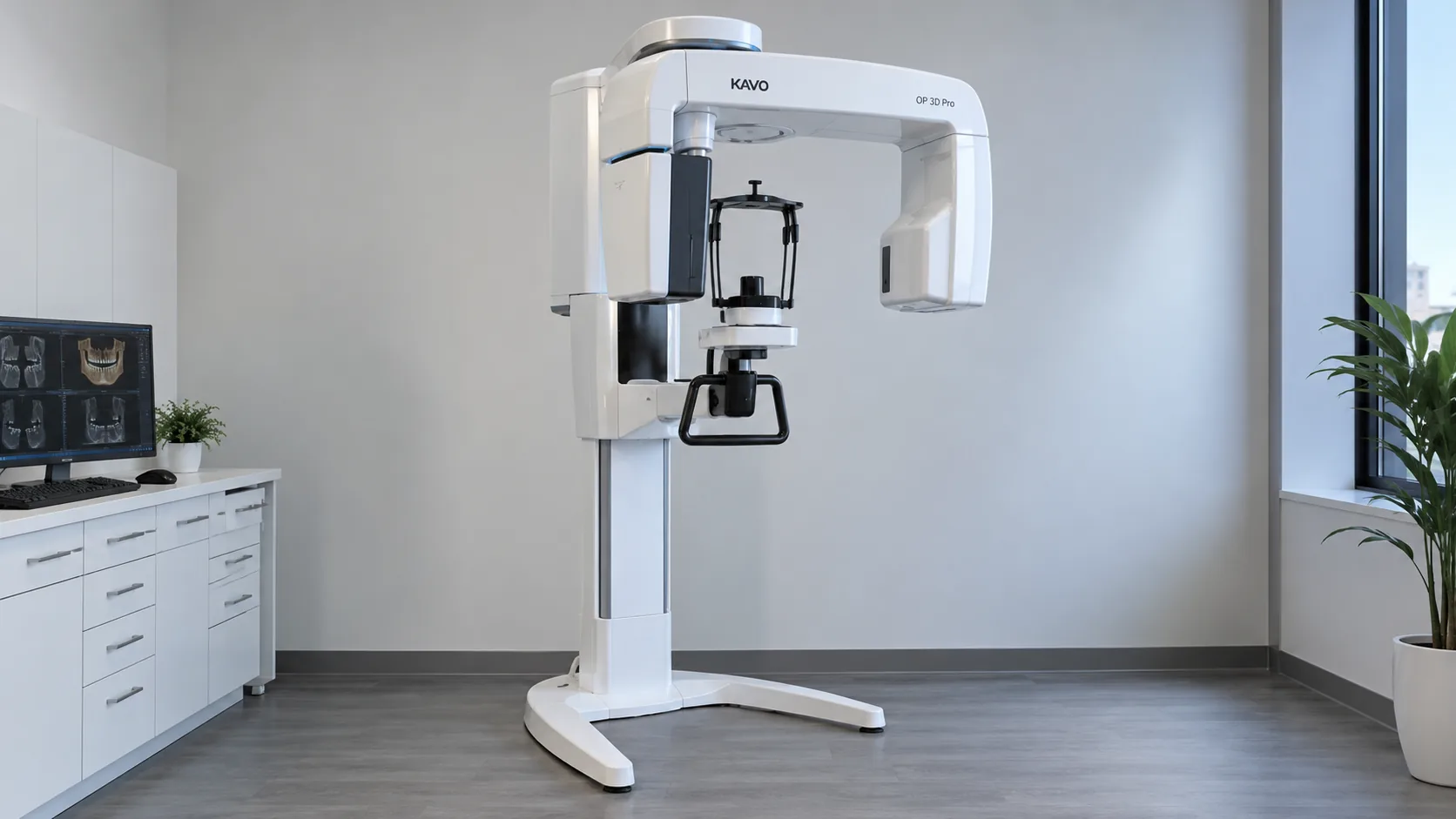
A cone beam CT scanner. One rotation, one three dimensional image.Fig. 01
A shadow, not a shape
A regular dental X-ray is a flat picture of something that isn’t flat. Everything between the beam and the sensor is stacked into one image, so a root, the bone in front of it and the bone behind it all land on top of each other.
01 · The instrumentOne scan, three planes
A cone beam CT (CBCT) scanner turns once around the head and builds a three dimensional image of the teeth, the jaw bone and the structures around them. Instead of one flat picture, the result is a volume that can be turned and sliced in any direction.
Each slice is read in three standard planes. Together they show what a flat X-ray can only hint at.
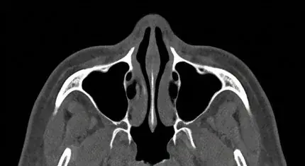 Axial · from above
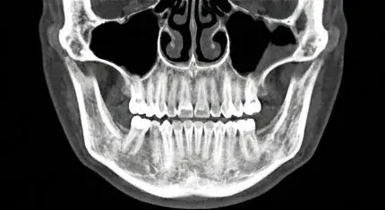 Coronal · from the front
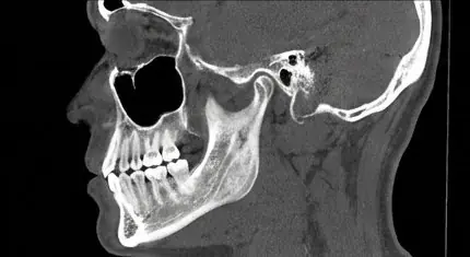 Sagittal · from the side
02 · What it showsWhat a flat image can’t show
- Structures overlap one another
- Height, but not width
- Distances can only be estimated
- Bone height and width, measured
- The nerve’s path through the jaw
- Sinus, jaw joints and airway
That matters most when a plan depends on exact measurements: where an implant can go and how long it can be, how close a root sits to a nerve, or what a jaw joint looks like before a bite is changed.
03 · The principlePlanned from what is actually there
Measuring first is the point of every instrument in the practice. With a cone beam scan, the decisions that depend on bone are worked out on the scan before treatment begins, not discovered during it.
“The decisions that depend on bone are worked out on the scan, not discovered during treatment.”
A cone beam scan uses more radiation than a single flat X-ray, so it isn’t a routine picture for every visit. It is taken when what it shows will change the plan. For implants, that is almost always: the next reading, Bone Before Implant, follows one scan through that planning.
04 · The seriesAbout this series
An ongoing series translating what the practice’s instruments actually reveal, one scan, one measurement, one plain-language explanation at a time.
- T-Scan: bite timingRead
- CBCT: bone in 3DYou’re reading
- JVA: joint soundSoon
- EMG: muscle effortSoon
- CEREC: fitSoon
- Waterlase: tissueRead
- Implants: bone firstRead
Words in this reading
CBCT
Cone beam computed tomography: a scanner that turns once around the head and builds a three dimensional image of the teeth, bone and joints.
Axial, coronal, sagittal
The three standard planes a scan is read in: from above, from the front and from the side.
Volume
The 3D image a cone beam scan produces, which can be turned and sliced in any direction rather than viewed as one flat picture.
Nerve canal
The channel through the lower jaw that carries the nerve giving feeling to the lower teeth, lip and chin.
Want to see what a scan would show in your case?
Book a visit at The Smile Galleries in Melbourne, Florida to talk through whether a cone beam scan would change your plan.
Dr. Sangiv I. Patel
Dr. Patel founded The Smile Galleries in Melbourne, Florida in 1995. Thirty years of Physiologic Dentistry: bite, joint, muscle and bone measured as one system before any treatment plan is written.
More about Dr. PatelNew readings, by email
Get the next reading when it’s published.
One plain-language article at a time, by email. No promotions, no schedule to keep up with.