- Home
- Technology
- Digital X-Rays & Imaging
Digital X-rays and imaging.
Most breakdown in the mouth happens where it can't be seen: between the teeth, under fillings, below the gum line and inside the bone. Digital X-rays let Dr. Patel see it, clearly.
- Radiation
- Up to 90% less than film
- On screen
- Right away, enlarged and adjusted
- Sharing
- Secure, in seconds
- In the practice
- Digital X-rays since 2007
Inside the mouth, and outside it.
X-rays are never taken on a calendar schedule. They're taken when a clinical question needs objective data, because in physiologic dentistry a treatment plan isn't made until the system underneath has been measured.
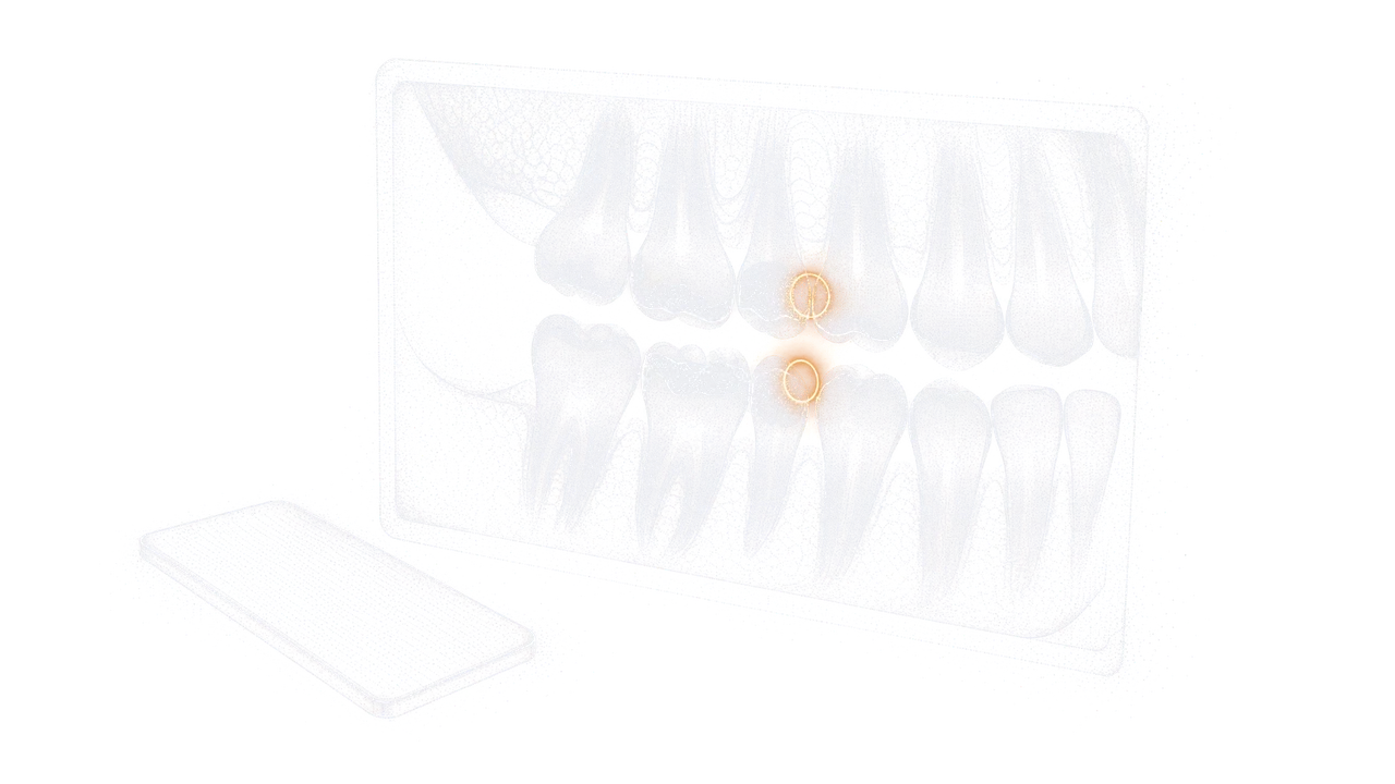
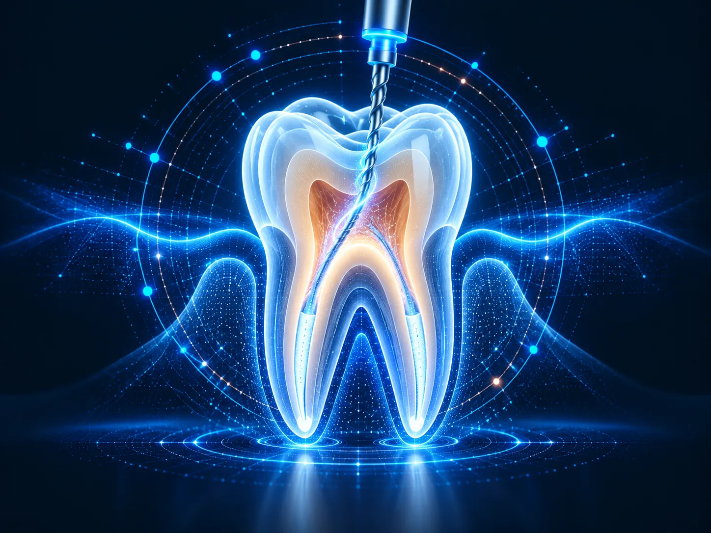
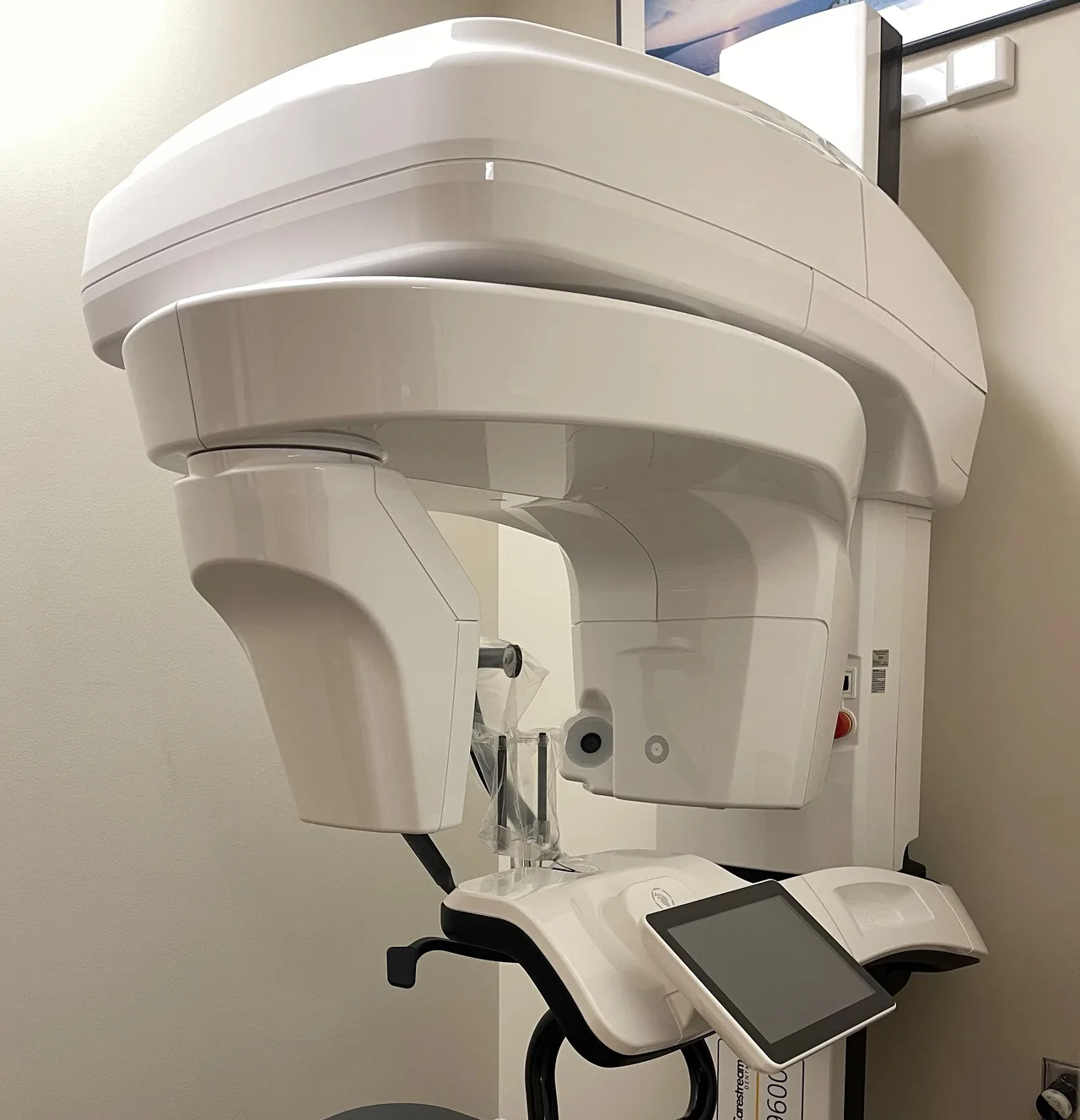
Between the teeth
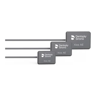
You bite on a small sensor. Shows decay between teeth, bone levels and how fillings and crowns fit.
 When more detail is needed
What a digital X-ray finds.
Hidden decay
Between the teeth, and under existing fillings and crowns.
Bone levels
Small changes in the bone that holds the teeth, to track gum health.
Roots
Root canal anatomy, nerve canals, and infections at the root tip.
Jaw
Lesions and cysts in the bone, found early.
Injuries
Root fractures and damage after trauma to the face.
Restorations
The fit at the edges of crowns, bridges and fillings.
TMJ
Bone changes that contribute to TMJ disorders.
- 90%
Up to 90% less radiation than traditional dental film.
Low dose, clean sensors.
Low dose
Under current ADA and FDA guidance, the dose from digital X-rays is low enough that lead aprons and thyroid collars aren't routinely needed. A lead apron is always available if you'd like one.
Clean sensors
Every sensor is covered by a fresh protective barrier, replaced after each patient.
Pregnant or nursing
Tell Dr. Patel first. Urgent imaging is safe when it's needed; anything that can wait is postponed until after delivery.
See what we see.
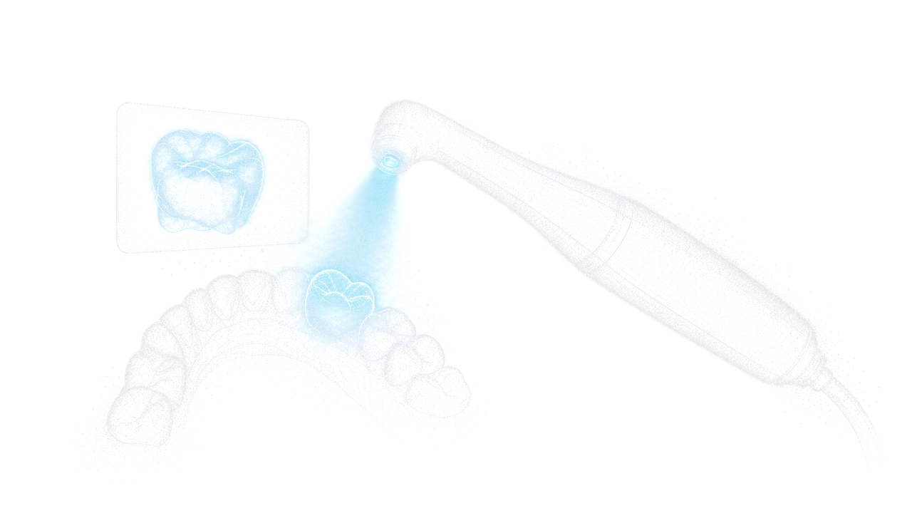
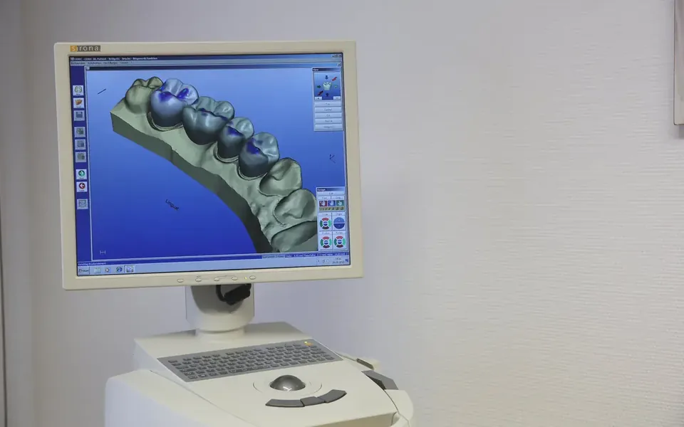
A pen-sized camera shows a live, high-definition view of your teeth on a screen, so you see exactly what Dr. Patel sees: hairline cracks, erosion and early decay. The images are saved and used again at the consultation and during treatment.
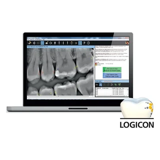
Decay detection, double-checked
Computer-aided software that reads digital X-rays to help confirm decay between the teeth in borderline cases, on natural and restored teeth, so the decision doesn't rest on a guess.
How often do I need dental X-rays?
There is no fixed schedule. How often depends on your risk of decay, your gum health, your age and what the exam finds. Dr. Patel orders X-rays only when a specific clinical question needs an objective answer.
Are digital X-rays safe?
Yes. They use up to 90% less radiation than traditional film. Under current ADA and FDA guidance the dose is low enough that lead aprons aren't required, but we keep them on hand for anyone who prefers one.
Can I have X-rays while pregnant?
If an urgent problem comes up, yes, with the proper precautions. If imaging can safely wait, we schedule it for after delivery. Please tell us before your visit if you are pregnant or nursing.
Why do I need X-rays if nothing hurts?
Decay, bone loss and root infections are usually silent at first. Finding them early means simpler, more conservative treatment, before symptoms start.
Can my X-rays be shared with other providers or my insurance?
Yes. Digital files can be sent securely in seconds to specialists, laboratories or your insurance company, so tests aren't repeated and approvals move faster.
Read more about what imaging shows.
 Read · 4 minA Jaw in Three Planes: Reading a CBCTA flat X-ray shows a shadow. A cone beam scan shows the teeth, the bone and the joints as a volume you can turn, slice and measure.Read the article
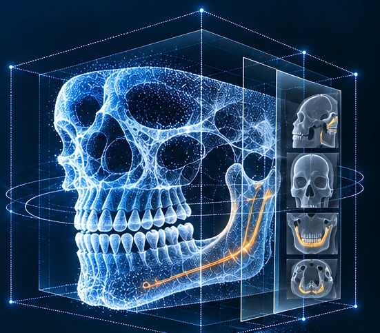 Read · 4 minBone Before Implant: Measuring the FoundationAn implant is only as steady as the bone it sits in. Height, width and the nerve's path are measured before anything is placed.Read the article
Want to see what an X-ray would show in your case?
Book an appointment to find out, or call us with any questions.